Today I’d like to focus on research performed by a group of physicians in São Paulo, Brazil, including Drs. Amato, Saucedo, Santos, and Benitti. Their paper is titled “Ultrasound criteria for lipedema diagnosis” and it was published in the medical journal Phlebology in the April 2021 issue. This study aimed to use ultrasound imaging to evaluate the thickness of the skin and underlying tissue in areas typically affected by lipedema, comparing women with and without lipedema. By doing this, it was their hope to develop skin and tissue thickness as a distinguishing feature of lipedema that could help with making an accurate diagnosis.
Methods
The study included 89 women, 63 of whom were diagnosed clinically with lipedema. A diagnosis was arrived at using a screening tool (The Lipedema Screening Questionnaire) developed by this same group of physicians in Brazil.
The criteria for diagnosis included onset after puberty; bilateral symmetrical fat deposit below the hip but sparing of feet; non-pitting edema in the legs that do not resolve with elevation; painful affected areas that are sensitive to pressure; and increased capillary fragility, with spontaneous bruising.
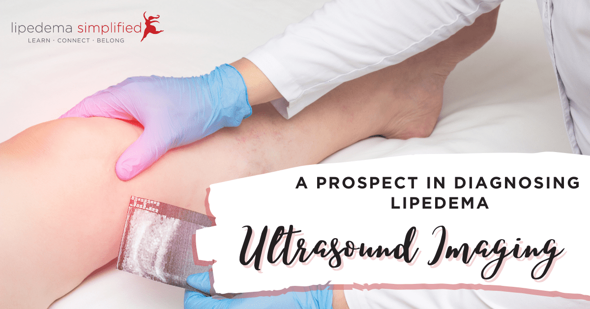
Ultrasound imaging was performed on all participants in predetermined spots including the front of the thigh, the front and outsides of the calf, and just above the inside of the ankle.
Study Participants
Here are the characteristics of the study participants:
The average age was 46 and the average BMI was just over 29 (overweight). 39 percent of participants were classified as obese (BMI>30) or morbidly obese (BMI>40) and 40 percent were overweight (BMI = 25-29). Only 20 percent of participants were considered to have normal weight.
There was a higher proportion of participants who were considered to be overweight and obese in the lipedema group. All of the women in this study also had evidence of venous disease, but any that were in an advanced stage were excluded from the study.
Finding a Distinguishing Feature
The authors wanted a distinguishing feature to help differentiate lipedema from its common comorbidities, such as venous disease and obesity. In their own clinics and in the literature, venous disease is present in 45 percent of women with lipedema, while obesity may be seen in up to 80 percent. The authors also state that the symptoms of lipedema can mimic some of those also present in venous diseases, such as a feeling of heaviness and swelling in the legs. Also adding to the difficulty in making an accurate diagnosis is that overall weight does not take into account fat distribution or body proportions, so this is not an effective measure for lipedema. Clearly, an effective and more accurate method to detect if lipedema is also present is needed.
Ultrasound
Ultrasound was used in a 2010 study to distinguish between lipedema and lymphedema and determined that the skin and underlying tissue were thicker in women with lipedema compared to those with lymphedema. Another earlier study also used ultrasound, but this time to stage the severity of lipedema depending on the amount of skin/tissue thickness present. For these reasons, the authors hoped to use the same technique to identify the existence of lipedema even if venous disease and/or obesity were also present.
Results
There was a statistically significant difference in skin and tissue thickness between those with and without lipedema in all of the body locations studied when the women were not grouped by BMI. If the BMI/weight classifications were used, only the area just above the inside ankle was not statistically significant. The authors suggested a minimal threshold or severity of thickness for each location could be used for diagnosing lipedema.
Takeaway
This study may have implications for physicians who want another method of confirming a diagnosis of lipedema. Many doctor’s offices have an ultrasound device with the sensitivity described in the article. This method is not invasive, painful, or expensive and has the added benefit of being quick and easy.
For more related content, be sure to check out Lipedema Simplified’s Flash Briefings, our daily mini-podcast with tips, tools, helpful research, and other resources pertaining to lipedema.
~ Leslyn Keith, OTD, CLT-LANA
Board President, Director of Research | The Lipedema Project